NOW WHAT? –
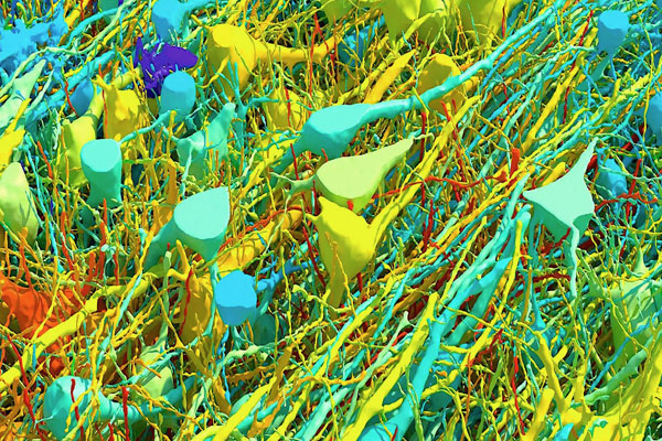
Aug. 15, 2024. – THIS IMAGE COULD be hung in a gallery, but it started life as a tiny chunk of a woman’s brain. In 2014, a woman undergoing surgery for epilepsy had a tiny chunk of her cerebral cortex removed. This cubic millimeter of tissue has allowed Harvard and Google researchers to produce the most detailed wiring diagram of the human brain that the world has ever seen.
Biologists and machine-learning experts spent 10 years building an interactive map of the brain tissue, which contains approximately 57,000 cells and 150 million synapses. It shows cells that wrap around themselves, pairs of cells that seem mirrored, and egg-shaped “objects” that, according to the research, defy categorization. This mind-blowingly complex diagram is expected to help drive forward scientific research, from understanding human neural circuits to potential treatments for disorders.
“If we map things at a very high resolution, see all the connections between different neurons, and analyze that at a large scale, we may be able to identify rules of wiring,” says Daniel Berger, one of the project’s lead researchers and a specialist in connectomics, which is the science of how individual neurons link to form functional networks. “